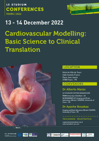
Cardiovascular Modelling: Basic Science to Clinical Translation

December 13, 2022 - December 14, 2022
Conference

Hôtel de Ville de Tours
Place Jean Jaurès
37000 TOURS
France

Presentation

Neurological and cardiovascular diseases are the leading cause of death globally. The use of computer models to simulate the functioning of the human body is viewed increasingly as one of the most promising tools to embrace and better understand the complexity of human pathophysiology, and therefore improve prevention, diagnosis, and treatment of human disease. A significant motivating factor for deployment of biomedical codes in clinical management of cardiovascular disease is the development of human digital twins. Such models would allow personalised guidance for healthcare, disease diagnosis and treatment, and wellbeing for specific individuals. The symposium will focus on characterization of blood flows using modelling as well as imaging tools (e.g., ultrasound) and its links to vessel wall mechanics and cardiovascular disease. This is an area that sees participation and expertise from different fields including vascular biology, fluid mechanics, computer science, medicine, but also imaging, medical device and software certification and regulation for translation into healthcare. The scientific literature in this field offers a rich spectrum of research, from basic science focusing on detailed mathematical, numerical and experimental descriptions of vascular pathophysiology, to applied research where similar approaches are used to test clinical hypotheses. The impact of such a tremendous, cross-disciplinary effort is starting to emerge, with such approaches starting to have an impact on healthcare. The event will aim to promote discussion and shine light on current and emerging research trends in the computational and experimental characterization and role of cardiovascular blood flows, arterial and venous wall mechanics, its correlation with vascular disease, with a specific emphasis on their application to unsolved clinical challenges and translation into healthcare.

Topics

  • Patient-specific, image-based modelling;
  • Reduced-order modelling;
  • Cerebral flow modelling;
  • Cerebral blood flow measurement;
  • Cardiovascular disease;
  • Cardiovascular physiology;
  • Clinical translation and applied research;

CONVENORS


Documents

Confirmed speakers

Click on the name to display the abstract

  • Dr Ayache Bouakaz, Imaging and Brain laboratory (iBrain) / INSERM, University of Tours
    Dr Ayache Bouakaz

    Imaging and Brain laboratory (iBrain) / INSERM, University of Tours

    10 boulevard Tonnellé 
    37000 Tours - FR

    Email: ayache.bouakaz@univ-tours.fr
    Tel.: +33247366142

    Ayache Bouakaz obtained his DEA degree (MSc) and his PhD in acoustics in 1992 and 1996 at the National Institute of Applied Sciences in Lyon, France (INSA Lyon). In 1998, he joined Pennsylvania State University at State College, PA, USA as a post-doc for 2 years. From December 1999 to November 2004, he held a position of associate professor at Erasmus Medical Center in Rotterdam, the Netherlands. His research focused on ultrasound imaging, ultrasound contrast agents and transducer design.

    In 2004, he obtained a position as an Inserm researchere (CR1) and since 2009, he has held the position of research director in the Inserm Imaging and Brain unit, where he heads the ultrasound imaging and therapy group. His research focuses on imaging and therapeutic applications of ultrasound.

    Ayache Bouakaz is a "chair professor" at the Jiaotong University of Xi'an in China since 2017. He is the general chair of the international conference IEEE International Ultrasonics Symposium (IEEE IUS) 2016 in Tours, France and co-general chair of the IEEE IUS 2021 edition and he was the vice-president of the IEEE UFFC society from 2017-2021.

    He has published more than 135 articles in peer-reviewed journals, more than 100 articles published in conference proceedings and has filed 9 patents.

  • Dr Alberto Marzo,
    Dr Alberto Marzo

    LE STUDIUM Visiting Researcher
    Insigneo (Institute for in Silico Medecine), University of Sheffield

    Department of Mechanical Engineering
    The University of Sheffield
    Pam Liversidge Building
    Mappin Street, Sheffield - UK

    Email:  a.marzo@sheffield.ac.uk
    Tel.: +447821196852
     

    Dr Marzo is an Associate Professor of Cardiovascular Biomechanics at the University of Sheffield, UK. 
    His research expertise is firmly rooted in Engineering and Mathematics in the context of 
    computational biomechanics, with a PhD on the mechanics of flow through elastic vessels from the 
    University of Sheffield, a David Crighton Research Fellowship at the University of Cambridge, and 
    participation in several EU/UK projects, focusing on cardiovascular flow characterisation and 
    development of computational frameworks. His expertise has a strong emphasis on clinical 
    interpretation and translation, with his time spent as Clinical Scientist for the UK National Health 
    Service. This experience gave AM exposure to the UK healthcare system, where he has witnessed the 
    often-difficult clinical perception of innovation, and the resulting challenges relating to technology 
    adoption.

    The role of collaterals in treatment of intracranial aneurysms
    Intracranial aneurysms are balloon-like focal deformations of a blood vessel in the brain, carrying an inherent risk of rupture and bleeding with severe consequences for the patient. A minimally invasive treatment option consists of deploying a medical device (Flow Diverting Stent or FDS) to trigger blood coagulation inside the aneurysmal sac to stop or prevent any existing or potential bleeding. In certain circumstances this treatment causes the occlusion of the arteries surrounding the aneurysm, leading to permanent and often irreversible damage (stroke). There is a strong consensus in the literature that the underlying causes might be associated with stent-induced adverse alterations to blood flow in the vascular network surrounding the aneurysm. Using a 3D-0D multidimensional patient-specific models of blood flow through an extended portion of the Circle of Willis, this study explores the possible role of collateral networks that are often associated with FDS-induced post-treatment complications. The long-term aim is that of providing guidance to clinicians towards a safer and more effective treatment of this condition.

  • Dr Ivan Benemerito,
    Dr Ivan Benemerito

    Institutions: The University of Sheffield

    Address: Sheffield, UK

    Email: i.benemerito@sheffield.ac.uk

    Phone: (+)

    Dr Benemerito is a research associate at The University of Sheffield and the INSIGNEO institute for in silico medicine. Following the award of his PhD from The University of Sheffield, during which he studied the contact mechanics of the ankle joint during walking activities, he shifted fields of application to cardiovascular biomechanics. He is interested in combining deterministic techniques with statistical approaches to investigate the sensitivity of complex computational models to uncertain input parameters, and to develop methodologies that can be translated to the clinical practice with minimum modifications to existing clinical practices.

    Identification of biomarkers for distal perfusion following an ischaemic event: a combined mechanistical-statistical approach

    An ischaemic stroke (IS) is an occlusion of a major cerebral vessel. It often causes severe reduction to the perfusion of downstream districts and the consequent death of tissues. The effects of middle cerebral artery (MCA) occlusion are partially mitigated by the action of the leptomeningeal anastomoses (LMAs), small arterioles which provide continuity of perfusion through diversion of flow from the anterior and posterior brain districts. Extensive LMAs networks are linked to better post-IS outcomes. The action of these vessels, which are active and can be observed only during an ischaemic event, induces modifications in the blood velocities of adjacent vessels. Transcranial Doppler ultrasound (TCD) can monitor the blood velocity in these major arteries but fails in detecting signals from the LMAs and to estimate the efficacy of their action in restoring distal perfusion. 1D computational models can simulate the cerebral circulation and identify biomarkers that correlate with the level of distal perfusion. In this study we have developed a deterministic1D model of the brain circulation which includes the LMAs, and combined its predictions with the use of Gaussian Process emulators and Sobol's sensitivity analysis to identify TCD biomarkers for distal perfusion in case of MCA stroke. We identified four biomarkers related to velocity pulsatility whose values can be associated with poor perfusion in the distal MCA regions.

  • Sara Bridio,
    Sara Bridio

    Politecnico di Milano

    Piazza Leonardo da Vinci 32 
    Milano - IT

    Email: sara.bridio@polimi.it
    Tel.: +390223993399

    I got my Master Degree in Biomedical Engineering at Politecnico di Milano in 2018. I’m currently a PhD candidate in Bioengineering at Politecnico di Milano, under the supervision of Prof. Francesco Migliavacca, and I will defend my thesis in early 2023. The topic of my research is the development of computational models of the treatments for stroke, from high-fidelity finite-elements models, to reduced order models. Part of my research was carried out within the EU Horizon2020 project INSIST (IN Silico trials for treatment of acute Ischemic STroke). In July 2022 I received the Young Researcher Prize at the European Solid Mechanics Conference in Galway (Ireland) with a work on the investigation of the impact of thrombus composition on the outcome of thrombectomy procedures.

    Dimensionality reduction and kernel optimization for the prediction of thrombectomy outcomes

    Endovascular thrombectomy (EVT) is the main treatment for acute ischemic stroke, aiming at removing the thrombus from a cerebral artery with a stent-retriever. The treatment must be performed in the first few hours from symptoms onset, allowing a short time window for pre-operative planning. Computational simulations of EVT can predict the treatment outcome but require a long computational time. In this work, a classification model is proposed, trained on high-fidelity EVT simulations, for providing fast estimates on the success of the procedure. The training dataset is composed of 94 finite-elements patient-specific EVT simulations, containing cases with successful (thrombus removed) and unsuccessful (thrombus not removed) outcomes, which are highly unbalanced: 81 successful against 13 unsuccessful EVTs. The proposed strategy is made of two steps. First, a dimensionality reduction approach is used to parametrize the vascular anatomies with few dimensions. Then, given as inputs the anatomy description and the thrombus properties for each virtual patient, a kernel function is trained, based on a restricted semidefinite positive optimization, with which clusters are created according to the EVT success. With the obtained tool, once data describing a new patient is available, it is processed through the trained kernel so that the results may approach either the successful or the unsuccessful cluster, suggesting a higher or lower probability of success of a possible EVT intervention.
     

  • Diederik Bulters ,
    Diederik Bulters

    University Hospital of Southampton

    Tremona Road
    Southampton SO16 6YD - UK

    Email: dbulters@uhs.nhs.uk
    Tel.: +447980596273

    Professor Bulters is a consultant neurosurgeon specialising in vascular conditions. His group’s main themes are risk prediction for unruptured intracranial aneurysms and reducing brain injury from haemoglobin after subarachnoid haemorrhage.
    He is past president of the British Neurovascular Group, has held grants from the NIHR, EPSRC, Innovate UK, MRC, European Union, RCS and several medical charities and has been chief and principal investigator for many randomised trials of new drugs, cell therapies and surgical techniques. 
    His interest in unruptured aneurysms focuses on risk prediction, new imaging techniques to stratify risk, including vessel wall imaging and dynamic contrast enhanced imaging, and the influence of modifiable risk factors including aspirin, blood pressure and cholesterol.

    Risk of Aneurysm Rupture study – rationale and opportunities

    Unruptured intracranial aneurysms (UIA) are common. However, only some subsequently rupture. Prophylactic treatment is possible but reserved for those with a rupture risk high enough to justify this. The problem is that our estimates of risk are not accurate enough, and do not stratify patients into sufficiently high and low risk groups, to base these treatment decisions on.
    The best natural history data comes from the PHASES study. This has not been validated, and given the heterogeneity in the populations, methods and biases of the constituent studies, there is a need to do so. There are also many predictors not considered in PHASES, and PHASES is based on short term follow up (mostly 1 year) with little data on long term rates relevant to patients. We therefore designed the Risk Of Aneurysm Rupture (ROAR) study with the aims to: 1) test the accuracy of PHASES, 2) evaluate additional predictors, 3) assess long-term rupture rates.
    ROAR is a longitudinal multicentre study that identifies patients with UIA in neurosurgery units and determines rupture events using national databases of hospital admissions and deaths. This design enables long term follow-up in a large cohort of 20,000 patients (11,000 to date). 
    In this presentation we will describe the design of ROAR and discuss the opportunities to identify different imaging markers of risk of rupture provided by a cohort that is expected to identify several hundred aneurysms with baseline MRA that subsequently rupture.
     

  • Gaetano Burriesci,
    Gaetano Burriesci

    University College London

    UCL Mechanical Engineering
    Torrington Place,
    London WC1E 7JE - UK

    Email: g.burriesci@ucl.ac.uk
    Tel.: +442076793922

    Gaetano is Chair of Bioengineering at UCL (UK) and Group Leader of Bioengineering at Ri.MED Foundation (Italy).  He studied Mechanical Engineering at the University of Palermo (Italy) and completed his PhD in biomechanics on a joint project between the same university and the University of Sheffield (UK), where he was then appointed Research Associate.  Subsequently, he joined SorinGroup (now Corcym) as Research Manager, before taking his position at UCL.  His main research interests lie in the field of cardiovascular engineering and medical devices development, where Gaetano has made major progresses that have found application into largely adopted therapeutic approaches.

    Design, development and preclinical validation of a novel transcatheter aortic valve concept

    This talk describes the design, development and preclinical assessment of a new polymeric aortic valve suitable for transcatheter implantation.  The device consists of three polymeric leaflets and an adaptive sealing cuff, supported by a fully retrievable self-expanding wire frame made from superelastic NiTi alloy.  A parametric design procedure based on numerical simulations was implemented to identify design parameters providing minimum stress levels and operating energy for the valve leaflets.  The wireframe was optimised to minimise the stress levels during valve delivery and provide adequate anchoring.  Valve prototypes were manufactured by thermomechanical processing of the NiTi wire and automated dip-coating of the polymeric components.  The hydrodynamic performances of the valves were assessed in a cardiac pulse duplicator, in compliance with the ISO5840-3 standard, and compared to two reference valves suitable for equivalent implantation ranges.  A valve prototype was implanted in orthotopic position of an acute ovine model, confirming retrievability, secure valve anchoring, adequate leaflets motion, and no interference of coronary flow or mitral valve function.
    The proposed valve system demonstrated excellent hydrodynamic performance with significant reduction in paravalvular leakage and the potential to mitigate complications related to imprecise valve positioning.  This new concept may offer a safer and more economical TAVI solution to a broader range of patients.
     

  • Emmanuelle Chaigneau,
    Emmanuelle Chaigneau

    Institut de la Vision / INSERM, Sorbonne University

    17 rue Moreau
    75012 Paris - FR

    Email: emmanuelle.chaigneau@inserm.fr
    Tel.:  +33153462611
     

    I graduated in 2001 from ESPCI-ParisTech, a french "grande ecole", providing a master-level multidisciplinary training with a major in physics.
    For my PhD I studied neurovascular coupling with 2-photon microscopy imaging and became a Doctor in Philosophy from Sorbonne University (Paris 6) in 2005.
    Then I worked as a post-doc research fellow, mainly in University College London, and I performed research and development both in microscopy and in neuroscience.
    In 2015 I got a permanent position at INSERM in Paris, and since I have been focusing at research and development of microscopy and modelling tools to study neurovascular coupling in the laboratory of Serge Charpak.  

    Investigation of functional hyperaemia with an experiment-based model of brain vasculature

    Functional hyperemia is the local increase in blood flow that occurs in response to local activation of neurons. In the olfactory bulb, functional hyperemia involves complex velocity changes resulting from vessel dilations at several levels of the vascular tree. Disentangling the effects of dilations occurring at each level is difficult as diameter changes cannot be selectively manipulated in vivo.  Experiment-based modelling can provide further insight in this question. 
    The vasculature was divided into four functional units according to the kinetics of the diameter changes of individual vessels. A four-level network was then developed by reducing all vessels in each of these physiological units into an equivalent tube. The diameter of each equivalent tube was set so that its resistance matches the resistance of the physiological unit. A formal mathematical model was developed to calculate the flow in each compartment as a function of the diameters of each compartment.
    In response to diameter changes whose kinetics followed experimental measurements, the model predicted RBC velocity dynamics in the compartments that were in line with the experimental results. Furthermore, the model allowed to manipulate the inputted diameter changes and test their impact on the velocity and flow changes. Our modeling faithfully shows that RBC velocity dynamics in the olfactory bulb results from the timing and relative changes of diameter that occur in the different vascular compartments.
     

CONVENORS


PROGRAMME

TUESDAY 13TH DECEMBER 2022

  • 8:30 Welcome coffee & registration
  • 9:00 Official opening – Alberto Marzo / Ayache Bouakaz (iBrain) + Sophie Gabillet (Le Studium)

SESSION 1: CEREBROVASCULAR MODELLING

  • 9:30 Emmanuelle Chaigneau - Investigation of functional hyperaemia with an experiment-based model of brain vasculature
  • 10:00 Ivan Benemerito - Identification of biomarkers for distal perfusion following an ischaemic event: a combined mechanistic-statistical approach. 
  • 10:20 Ahmet Sen - Understanding the relationship between anatomical variations of the circle of Willis and hemodynamics using machine learning
  • 10:40 Weiquiang Liu - 3D blood flow simulations for understanding cerebral vasculopathy in sickle cell patients
  • 11:00 Coffee break

SESSION 2: CARDIOVASCULAR MODELLING AND MEDICAL DEVICES

  • 11:30 Francesco Migliavacca - Moving Aorta: from bench tests to bed side. 
  • 12:00 Benjamin Csippa - Measurement-supported computational framework for the hemodynamic investigation of flow diverter treatments. 
  • 12:20 Anna Ramella - On the modelling of the TEVAR procedure: a detailed FEA-FSI methodology. 
  • 12:40 Sara Bridio - Dimensionality reduction and kernel optimization for the prediction of thrombectomy outcomes
  • 13:00 Lunch (Hotel de Ville)

SESSION 3: ULTRASOUND APPROACHES IN CARDIOVASCULAR APPLICATIONS 

  • 14:30 Clément Papadacci - 3D ultrafast ultrasound applications for human heart characterization.  
  • 14:55 Guillaume Lacoin - Intraoperative ultrasound plane wave Doppler imaging allows better definition of gliomal infiltration. 
  • 15:15 Philippe Trochet - Innovative imaging for cardiovascular diseases. 
  • 15:40 Redouane Ternifi - Super-Resolution Ultrasound Imaging: Current Research and Applications
  • 16:05 Coffee break

SESSION 4: CLINICAL PERSPECTIVES AND THE ROLE OF IN SILICO MEDICINE 

  • 16:30-17:00 Ana Paula Narata - Imaging to create virtual humans: the importance of models to improve the management of vascular pathologies 
  • 17:00-17:30 Alfons Hoekstra – Towards In-Silico Stroke Trials
  • 17:30-18:00 Diederik Bulters - Risk of Aneurysm Rupture study – rationale and opportunities. 
  • 18:00 Closing remarks
  • 18:30 Public lecture in French: Dr Jean-Michel Escoffre & Prof. Damien Lacroix - Innover aujourd’hui pour guérir demain: diagnostics et thérapies personnalisés
  • 20:00 Social dinner

WEDNESDAY 14TH DECEMBER 2022

  • 8:30 Welcome coffee
  • 9:00  Welcome – Alberto Marzo / Ayache Bouakaz 

SESSION 5: CARDIAC MODELLING APPLICATIONS

  • 9:15 Gaetano Burriesci - Design, development and preclinical validation of a novel transcatheter aortic valve concept
  • 9:45 Nada Ghorab - Modelling Coronary Bifurcations: Using angiograms to produce CFD models that predict blood flow – the methodology.
  • 10:05 Giulia Pederzani - Digital Coronary Phantoms as Gold Standard Method for Software Validation and Improvement. 
  • 10:25 Pjotr Hilhorst - An in Silico clinical trial on coronary fractional flow reserve as a replacement for the original clinical trial: a feasibility study. 
  • 10:45 Coffee break

SESSION 6: CARDIOVASCULAR MODELLING

  • 11:15 Alberto Marzo - The role of collaterals in treatment of intracranial aneurysms. 
  • 11:35 Gabor Zavodszky - Components of the high-risk thrombotic blood flow environment, from cells to organs.
  • 11:55 Ning Wang - Determining image accessible biomarkers for non-invasively distinguishing hypertensive from diabetic renal injury through a mechanistic model and MR imaging. 
  • 12:15 Thomas Feaugas - Design of artificial vascular devices: hemodynamic evaluation of shear-induced thrombogenicity. 
  • 12:35 Closing lunch (Hotel de Ville)


Hôtel de Ville de Tours (Tours City Hall) 

Boulevard Heurteloup - 37000 TOURS - FR

The event will take place in the city centre at the Hotel de Ville of Tours located in Square Jean-Jaures and very near the main train station. This elegant building was designed in the early 20th century by Victor Laloux, the architect of the Gare d'Orsay in Paris (now the Musée d'Orsay).  Its monumental stairs, paintings and sculptures make it a recommended place to visit. Participants will also have the opportunity to discover French cuisine and Loire Valley wines during an evening social.

Partners of the event