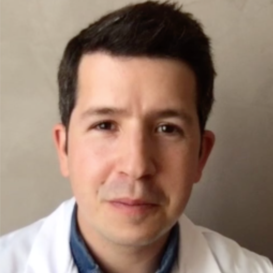
Pr Grégoire Boulouis is a clinician-scientist and interventional neuroradiologist at CHU Tours, France, affiliated with INSERM’s iBrain research unit (UMR1253) and CIC IT 1415. His expertise centers on adult and pediatric stroke. Trained in Lille, Paris, and at Harvard Medical School's Stroke Research Center, he actively participates in national and international stroke trials and leads multiple clinical research initiatives. A committed educator, he co-founded and chaired JENI, a collaborative research network for junior neuroradiologists. He holds leadership roles in the French Society of Neuroradiology, the International Pediatric Stroke Organization and the European Stroke Organisation, contributes to national and international clinical trials. With over 300 peer-reviewed publications, his research focuses on translating neuroimaging advances into improved patient outcomes.
The variety of vascular malformations, 3D visualization/models
Vascular malformations are complex anomalies requiring precise anatomical characterization for effective intervention. This talk explores the added value of 3D Cone Beam Computed Tomography and advanced visualization techniques, particularly cinematic rendering, for guiding direct puncture approaches in treating superficial vascular malformations.
In low-flow vascular malformations, needle placement is typically guided visually or by ultrasound, with subsequent phlebography or cystography to characterize lesion anatomy and monitor sclerosant injection. High-flow malformations present different challenges, with imaging revealing rapid arteriovenous shunts and dilated draining veins. Advanced imaging, including rotational 3D angiography, significantly aids therapeutic targeting. Sclerosants are administered through transarterial, transvenous, or direct puncture approaches, supported by precise anatomical mapping provided by CBCT (Cone Beam Computed Tomography).
In a recent work, we showed that CBCT significantly improved understanding of lesion architecture in 89.2% of cases and facilitated needle placement in 64.9% of interventions, particularly for complex cervical and ENT lesions.
These findings highlight the clinical relevance of integrating 3D visualization technologies such as CBCT and cinematic rendering into routine interventional radiology practice, ultimately translating into enhanced patient outcomes and communication. Imaging exemple will be presented and discussed.

Clinical Investigation Centre of Tours - Technological Innovation, Regional University Hospital in Tours
& Imaging, Brain, Neuropsychiatry (iBraiN) / INSERM, University of Tours
Address: Faculté de Médecine, 10 Boulevard Tonnellé 37032 Tours - France
Email: g.boulouis@chu-tours.fr
