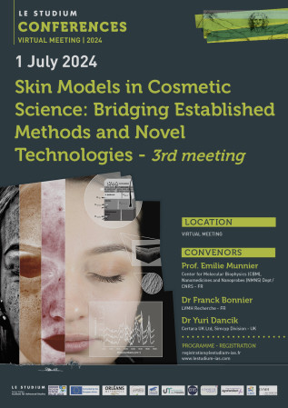
Skin Models in Cosmetic Science: Bridging Established Methods and Novel Technologies
3rd meeting

July 01, 2024 - 13 h 45 / 17 h 00
Conference
 Virtual meeting

Presentation


THIS EVENT IS REPLACED BY A FREE VIRTUAL MEETING ON 1st OF JULY 2024

LE STUDIUM is thrilled to announce the third edition of 'Skin Models in Cosmetic Science: Bridging Established Methods and Novel Technologies,' which will be held online as a free webinar. The upcoming webinar features speakers addressing the exploration and characterization of human skin in the dermo-cosmetic field, aiming to unite global experts to unravel the complexities associated with non-invasive analysis and strengthen transversal approaches across in vitro, in vivo, and in silico methods

Navigating the intricacies of in vivo analysis remains a paramount challenge in dermo-cosmetic research. Traditional techniques often grapple with ethical considerations, individual variability, and the inherent limitations of real-time measurements. In contrast, in vitro and ex vivo models provide a more controlled environment for researchers to scrutinize specific aspects of skin physiology, offering reproducibility and precision in experimentation. By simulating the intricacies of human skin, in vitro and ex vivo models become invaluable tools for researchers seeking a deeper understanding of physiological mechanisms and/or aiming to assess overall skin health. Emerging mathematical models provide predictions that allow interpretation of these data and guide further experimentation.   

The synergy between dermatology and skincare research goes beyond disorder management, contributing to studies on maintaining healthy skin. This dual impact fosters a holistic approach to skincare, where scientific advancements improve patient outcomes and empower individuals to achieve optimal skin health. The conference will act as a catalyst for collaboration, urging a harmonious alliance between academia, industry, and regulatory bodies. By forging interdisciplinary partnerships, participants can expect to expedite the translation of research findings into tangible applications. Moreover, the event will serve as a forum to explore future directions of dermo-cosmetic research, presenting a roadmap for innovative technologies, methodologies, and strategies that will shape the industry's trajectory. 

The conference will offer a platform for experts to discuss and share insights on the various challenges that involve the combination of biology, biophysics, mathematics, and analytical methods for advancing skin science: 

- New alternative methodologies (NAMs) and emerging analytical tools: In vitro, in chemico and in silico NAMs allow researchers to study the relationships between skin biology, skin barrier and the absorption of dermo-cosmetic topical actives. Of increasing interest in the cosmetic sciences, physiologically based pharmaco- or toxicokinetic models (PBPK or PBTK) can predict the absorption and disposition of cosmetics by integrating skin physiological data and physico-chemical data on the active and the formulation within a sound mathematical framework. They can help to interpret experimental data, guide in vitro experiments and in vivo trials and understand the impact of biological variability on product efficacy and safety.   

- Advancements and strategies in optimization of reconstructed skin models for in vitro testing: Nowadays, the challenge of developing and optimizing models that accurately mirror human skin architecture has led to sophisticated 3D models, pushing the boundaries for experiments on metabolically and functionally active human skin models and improving the effectiveness of in vitro testing. 

- Human skin explants to bridge the gap between in vitro and in vivo: Human skin explants remain widely used for ex vivo experimentation. Introducing the use of living skin explants further emphasizes the exploration of the dynamic properties and responses of skin tissues in conditions that mimic in vivo environments. Positioned at the intersection of in vitro and in vivo experimentation, a current challenge is to provide comprehensive results that support clinical studies by avoiding intrusive procedures to preserve the skin in its native state, thereby overcoming traditional invasive protocols. 

- In vivo skin analysis, a multimodal dilemma: Efficiently gathering valuable information from the skin, whether at the surface or deeper layers, hinges on the characteristics of the chosen technique. Recent strides in topographic, tomographic, biomechanical, and chemical assessment methods represent significant progress in refining the precision and depth of in vivo skin assessment. These advancements not only enable a more comprehensive understanding of skin physiology and skin health but also open new avenues for exploring the efficacy of topical products. 

- Recent innovation in approaches for dermo-cosmetic product efficacy and safety testing: Advancements in dermatological analytical tools, ranging from imaging devices to molecular diagnostics, facilitate the exploration of healthy skin; enable precise diagnosis and support the development of targeted treatments for conditions such as eczema and psoriasis. The accessibility and refinement of these technologies empower dermatologists and pharmacologists to unravel the complexities of skin health and treatment. Simultaneously, the skincare industry benefits from personalized solutions, utilizing cutting-edge methods for tailored treatments. 

This international conference is organised in the framework of the COSMETOSCIENCES ARD CVL Programme.

Convenors

Prof. Emilie Munnier
Center for Molecular Biophysics (CBM), Nanomedicines and Nanoprobes (NMNS) Dept / CNRS - FR

Dr Franck Bonnier
LVMH Recherche - FR

Dr Yuri Dancik
Certara UK Ltd, Simcyp Division - UK

 

Confirmed speakers

Click on the name to display the abstract

  • Dr Muriel Cario, Bordeaux Institute of Oncology (BRIC) / INSERM, University of Bordeaux - FR
    Dr Muriel Cario

    Bordeaux Institute of Oncology (BRIC) / INSERM, University of Bordeaux

    Address: 2 rue Dr Hoffmann Martinot , 3076 Bordeaux Cedex, France

    Muriel Cario currently works at INSERM 1312, University of Bordeaux.  She has over 25 years' experience in the physiopathology of human skin pigmentation and tissue engineering. She is a board member of the European Society for Pigment Cell Research (ESPCR) and of the Cosm'actifs GDR.  She collaborates with academic teams and the cosmetics industry to develop new models for studying skin pathologies such as senile lentigo, melasma, vitiligo, melanoma and carcinoma, new methods for vectorizing active ingredients and to test new active molecules. She has published over 70 articles and chapters on skin models, their use in deciphering skin diseases and testing active ingredients. Dr Cario is co-director of Aquiderm, a technology transfer unit specialized in dermocosmetic products.

    Skin models use to test active ingredients

    The skin is a complex organ composed of 3 layers (epidermis, dermis and hypodermis) which enable it to perform its various functions: defense against external aggression, thermoregulation and vitamin D production. These layers are made up of cells with very specific properties and roles, such as keratinocytes, which ensure the structure of the epidermis, melanocytes, which produce melanin, the skin pigment, and dermal fibroblasts, which produce collagen and elastin. Skin characteristics differ according to age, photo-exposure and phototype (skin color). In cosmetics, only in vitro and ex vivo models can be used; animal experimentation is no longer authorized. There are various models of varying complexity, from simple models made with keratinocytes on polycarbonate filters to skin explants. Skin models can be produced on acellular or cellular supports, with one or more cells from the dermis and epidermis. They can also be chemically stimulated or genetically modified to reproduce various skin alterations such as age spots (senile lentigo). The choice of the right model depends on a number of parameters, such as the target of the molecule, the time required to obtain an effect and the markers studied. For example, to study a molecule active on age spots due to pigment accumulation in the epidermal basal layer, a model composed solely of keratinocytes and melanocytes is not necessarily suitable. In this presentation, we will review the different 3D models and their advantages.

  • Prof. Arnaud Dubois, Optics Institute, University Paris-Saclay - FR
    Prof. Arnaud Dubois

    Optics Institute, University Paris-Saclay

    Address: 2 avenue Augustin Fresnel, 91127 Palaiseau cedex, France

    Arnaud Dubois received his Ph.D. in physics from Paris-Saclay university in 1997. Since 2006, he has been a professor of optics at Institut d'Optique in Palaiseau, France. His teaching activities encompass most aspects of optics at the master’s level. His research interests lie in the field of biomedical imaging. As a pioneer in optical coherence microscopy in the early 2000s, A. Dubois has since been a major contributor to the development of this technology. He has published 175 research articles and 12 book chapters and has participated in approximately 250 conferences. He has 5 patents to his credit. In 2014, he co-founded DAMAE Medical, a startup company engaged in the development of an innovative optical coherence microscopy technique for high-resolution skin imaging. 

    Skin imaging and characterization using line-field confocal optical coherence tomography (LC-OCT)

    Line-field confocal optical coherence tomography (LC-OCT) is an optical imaging technique based on a combination of the principles of optical coherence tomography (OCT) and reflectance confocal microscopy (RCM), with line-field illumination with broadband light and detection with a line-camera. LC-OCT can provide high-resolution (~1 μm) images of biological tissues non-invasively and in vivo. The value of the technique has been demonstrated particularly in dermatology and in cosmetology, thanks to its ability to generate cell-resolved images of the skin. This talk presents the LC-OCT technique and its application in dermatology and in cosmetology. The principle behind the technique is described, form its first developments in laboratory to the latest advances industrialized and commercialized by DAMAE medical. The technology has been miniaturized to fit within an ergonomic handheld probe, allowing for the easy access of any skin area on the body. A video camera was incorporated into a handheld probe to acquire dermoscopic images in parallel with LC-OCT images. A confocal Raman spectrometer was associated with a LC-OCT device to record morphological images of the skin in which points of interest can be subjected to molecular characterization. Dedicated algorithms based on artificial intelligence have been developed to automate the segmentation of various skin structures and for the computation of quantification metrics. 

  • Dr Hichem Kichou, Center for Molecular Biophysics (CBM), Nanomedicines and Nanoprobes (NMNS) Dept / CNRS - FR
    Dr Hichem Kichou

    Center for Molecular Biophysics (CBM), Nanomedicines and Nanoprobes (NMNS) Dept / CNRS 

    Address: 31 Avenue Monge 37200 Tours, France 

    Email: Hichem.kichou@univ-tours.fr

    Hichem KICHOU holds a PhD in analytical chemistry from the University of Tours, France. His doctoral research focused on developing comprehensive multi-method analytical approaches to study the interaction and diffusion of active ingredients, both cosmetic and pharmaceutical, within human skin. His work involved using advanced chromatography techniques to investigate the penetration kinetics of molecules across different skin layers. Additionally, he used Raman spectroscopy combined with multivariate analysis to explore skin penetration profiles. This innovative methodology provided significant insights into the behaviour of active ingredients in dermatological applications, advancing scientific knowledge and practical applications in skin research. 

    Exploring Reconstructed Human Skin and Synthetic Membranes as Alternatives to In Vitro Permeation Testing.

    In the development and optimisation of dermatological products, in vitro permeation tests (IVPT) are essential for studying skin penetration. To improve standardisation and replicate the properties of human skin, several models are explored as alternatives. Among these models, Strat-M® and RHE models stand out. Strat-M®, a polymer model, does not replicate the formulation effect observed on human skin. However, it shows permeability closer to human skin compared to RHE. RHE, while reproducing the formulation effect and demonstrating qualitative molecular similarity to human skin through Mass Spectrometry and Raman Spectroscopy (RS), shows higher permeability values than human skin. This makes it a good alternative for screening formulations, highlighting its importance as a reliable substitute. However, a major challenge with RHE is its limited shelf life. To address this, the chemical fixation of RHE in formalin for 24 hours has been examined for storing samples for up to 21 days. RS analysis revealed that while fixation alters the biochemical architecture, particularly proteins and lipids, it does not significantly affect the cumulative amount and permeability in IVPT using caffeine as a model compound. This preservation method offers increased flexibility and utility in skin model research, opening up prospects for mitigating the storage limitations of RHE models while maintaining their performance as effective barriers for rate-limiting diffusion of active molecules.

  • Dr Sebastian Polak, Certara - UK / Jagiellonian University - PL
    Dr Sebastian Polak

    Certara UK, Simcyp Division

    Address: Level 2, Acero, 1 Concourse Way, Sheffield S1 2BJ, UK

    Faculty of Pharmacy, Medical College, Jagiellonian University

    Address: Medyczna 9 Street, 30-688 Kraków, Poland

    Email: sebastian.polak@certara.com

    Dr Sebastian Polak is also a Senior Scientific Advisor in Certara UK, part of an international Certara company leading the team developing non-oral in silico absorption models. Dr Polak also holds tenure position at the Faculty of Pharmacy Jagiellonian University Medical College, Krakow, Poland (Professor of Biopharmacy) where he leads a multidisciplinary team of scientists and engineers working on applying various modelling and simulation approaches in drug development.
    Always late, lacks assertiveness, likes motorcycles theoretically and even more in real life.

    Utilization of PBPK models is a standard approach in the pharmaceutical industry realm, so why is cosmetics industry afraid of using this tool effectively for its benefit?

    In vitro modelling and simulation using Physiologically Based Pharmacokinetic (PBPK) modelling has become an important part of the drug development process, both for novel and generic drug products [1]. Among others USFDA supports the use of modelling and simulation to assess: DDI risk, special population PK, bioequivalence between reference and test products [2]. Incorporation of mechanistic biophysically detailed model describing the behaviour of various semisolid formulations after application on the skin allows one to account for critical quality attributes (CQAs) and potentially optimize the formulation [3,4]. Hundreds of individual label claims have been approved based – at least partially – on the results of PBPK simulations with clinical trials waived. Utilization of PBPK/PBK models in the cosmetic industry seems to be limited [5]. My aim is to discuss this matter and together with the audience define potential reasons for this situation. I also hope to define routes of expansion of modelling and simulation-based approaches in the field of dermatokinetics assessment and formulation optimization of cosmetics.
    References: (1) Shebley M. et al. Clin Pharmacol Ther. 2018 Jul;104(1):88-110. doi: 10.1002/cpt.1013 (2) Tsakalozou E, et al. AAPS J. 2023 Oct 2;25(6):96; doi:10.1208/s12248-023-00862-x. (3) Patel N et al. CPT PSP. 2022 Aug;11(8):1060-1084; doi:10.1002/psp4.12814. (4) Shah H, et al. Pharmaceutics. 2023 Mar 23;15(4):1040. doi: 10.3390/pharmaceutics15041040. (5) Krstevska A, et al. Pharmaceutics. 2022 Dec 28;15(1):107. doi: 10.3390/pharmaceutics15010107.

  • Dr Gerwin Puppels, RiverD international B.V. - NL
    Dr Gerwin Puppels

     RiverD international B.V.

    Address: Marconistraat 16, 3029 AK Rotterdam, Netherlands 
     Email: gpuppels@riverd.com 

    Gerwin Puppels is the founder and managing director of RiverD International (www.riverd.com), which develops instruments and diagnostic applications, based on Raman spectroscopic tissue e analysis. An example is the gen2-SCA family of in vivo skin analysis instruments. He is an associate professor at the Erasmus-university Medical Center in Rotterdam, The Netherlands and has been active in the field of Raman spectroscopy for over 30 years, making significant contributions to the development of the technique. His current research interests include skin analysis and Raman-guided surgery/biopsy. He has authored over 150 peer-reviewed papers and 18 patents. He holds a Ph.D in applied physics from the University of Twente, The Netherlands.

    Skin models vs in vivo skin: insights from Raman spectroscopy

    Skin models play an important role in the transition to animal free testing. However, just as animal skin is not the same as human skin, skin models are not human skin, even if they look much the same under a microscope. One of the important functions of the skin is barrier formation; to keep water in and to keep the outside world … out. That process starts immediately after birth. In vivo Raman spectroscopy offers a clear, non-invasive window on this barrier development. 
    The presentation will summarize the information about the skins’ molecular composition and molecular anatomy, barrier formation and skin penetration, that can be obtained by in vivo Raman spectroscopy. 
    It will then make the comparison between in vivo skin and various skin models with respect to these aspects. Raman spectroscopy could serve as a great, perhaps sometimes confronting, tool to further guide and test the development of skin models towards becoming real life-like skin models, in appearance, but also in terms of molecular composition and skin physiological and functional aspects. 

  • Dr Dandan Tu, Wellman Center for Photomedicine at Massachusetts General Hospital & Harvard Medical School - USA
    Dr Dandan Tu

     Wellman Center for Photomedicine at Massachusetts General Hospital & Harvard Medical School 

    Address: CNY149-3, 13th St, Charlestown, 02129, MA, USA 

    Email:  datu@mgh.harvard.edu

    Dr Tu is currently a postdoctoral research fellow in Prof. Conor Evans’ lab at the Wellman Center for Photomedicine at Massachusetts General Hospital & Harvard Medical School. She received her Ph.D. from the Biomedical Engineering Department at Texas A&M University in 2021. During her doctoral research, she specialized in using Raman spectroscopy to detect biomarkers in biological fluids. Her current research focuses on coherent Raman imaging, specifically Stimulated Raman Scattering (SRS), and its applications in studying the pharmacokinetics and pharmacodynamics of topical dermatological formulations.

    Determining topical product bioequivalence with stimulated Raman scattering microscopy.

    Stimulated Raman scattering (SRS) microscopy has unique capabilities enabling continuous, high spatial and temporal resolution and quantitative imaging of drugs within the skin. In this presentation, we will use an example to show its application in studying of the topical dermatological formulations. Specifically, we developed an approach based on SRS and a polymer-based standard reference for the evaluation of topical product bioequivalence (BE) in human skin ex vivo. BE assessment of tazarotene-containing formulations was achieved using cPK parameters obtained within different skin microstructures. The establishment of BE between the RLD and an approved generic product was successfully demonstrated. Another formulation containing polyethylene glycol as the vehicle was demonstrated to be not bioequivalent to the RLD. Compared to using the SRS approach without a standard reference, the developed approach enabled more consistent and reproducible results, which is crucial in BE assessment. The abundant information from the developed approach can help to systematically identify key areas of study design that will enable a better comparison of topical products and support an assessment of BE.

Programme

Monday 1st July 2024                                   


Video
Click on the title to display the video


Partners of the event