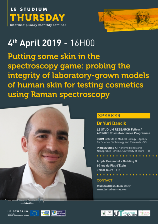
Dr Yuri Dancik

Établissement d'origine
Institut de biologie médicale - Agence pour la science, la technologie et la recherche - SG
Laboratoire d'accueil
Nanomédicaments et Nanosondes (MNMS), Université de Tours - FR
Hôte scientifique
Dr Franck Bonnier
PROJET
Molecular imaging using Raman spectroscopy: from fundamental research to industrial applications
We are using Raman spectroscopy to demonstrate the impact of storage conditions on the quality of lab-grown human skin equivalent tissues. The skin equivalents they are using are known as reconstructed human epidermis or RHE (Figure 1), as they mimic the uppermost epidermal layer of human skin.
The development, characterization and use of RHE is an active area of cosmetic and pharmaceutical R&D. Designed to replace animal tissues, RHE are particularly useful for testing or screening new cosmetic formulations and pharmaceutical topicals. To date, little is known on the effects of common tissue storage conditions on the quality and in particular, the barrier function, of RHE. Commercial RHEs are frequently cultured and shipped in batches of 6 or 12 replicates (Figure 1A). With testing and screening applications often requiring large numbers of replicates, practical knowledge on the effects of storage conditions is of significant value to academic and industrial RHE users.
The specific scientific goals are thus:
- To investigate the impact of different storage conditions on the barrier function of RHE. To this end, the penetration of a chemical applied to the RHEs stored under the different conditions is tracked via Raman spectroscopy (Figure 2A).
- To compare the chemical penetration results obtained via Raman spectroscopy to results obtained in parallel from a widely-used, but more laborious, methodology for studying skin penetration (Figure 2B).
- To assess how the information obtained by Raman spectroscopy complements that attained from the conventional skin penetration experiment.
In addition to answering a practical scientific question, the project is meant to highlight a novel application of Raman spectroscopy within the field of skin analysis.
Thus far we have optimized of skin penetration experiments and spectroscopic data acquisition through rigorous preliminary experimentation and the methodologies for data analysis, designed and performed a full experiment involving RHE replicates stored under various conditions, with both Raman spectroscopic and conventional skin penetration data obtained and analysed of the full experiment’s results.
The results obtained thus far are very encouraging and enable us to design follow-up experiments to fine-tune and complement our latest work.
Publications
Final reports
Reconstructed human epidermis (RHE) is an emerging skin model in pharmaceutical, toxicological and cosmetic sciences, yielding scientific and ethical advantages. RHEs remain costly, however, due to consumables and time required for their culture and a short shelf-life. Storing, i.e., freezing RHE could help reduce costs but little is known on the effects of freezing on the barrier function of RHE. We studied such effects using commercial EpiSkin™ RHE stored at −20, −80 and −150 °C for 1 and 10 weeks. We acquired intrinsic Raman spectra in the stratum corneum (SC) of the RHEs as well as spectra obtained following topical application of resorcinol in an aqueous solution. In parallel, we quantified the effects of freezing on the permeation kinetics of resorcinol from time-dependent permeation experiments. Principal component analyses discriminated the intrinsic SC spectra and the spectra of resorcinol-containing RHEs, in each case on the basis of the freezing conditions. Permeation of resorcinol through the frozen RHE increased 3- to 6-fold compared to fresh RHE, with the strongest effect obtained from freezing at −20 °C for 10 weeks. Due to the extensive optimization and standardization of EpiSkin™ RHE, the effects observed in our work may be expected to be more pronounced with other RHEs.


